
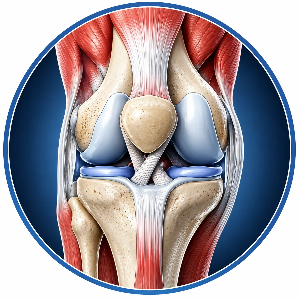
The knee joint is one of the largest and most important joints of the human body. It connects the femur (thigh bone) with the tibia (shin bone) and also includes the patella (kneecap).
The knee is mainly responsible and straightening the leg
- Providing stability during standing and walking
- Helping with running, jumping, climbing stairs and squatting
- Transferring forces between the hip and ankle
The knee is primarily a hinge-type synovial joint, although a small amount of rotation also occurs, particularly when the knee is flexed.
1. Bones Forming the Knee Joint
The knee consists of three main bones:
1. Femur
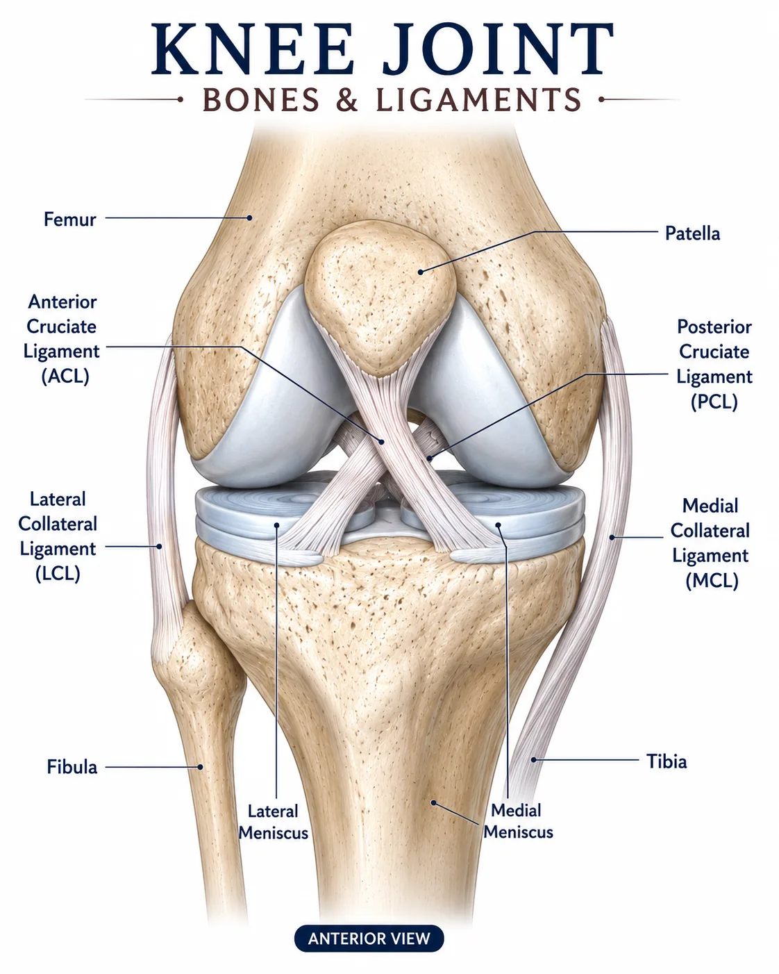
The femur is the thigh bone. Its lower end forms the medial and lateral femoral condyles, which articulate with the tibia.
2. Tibia
The tibia is the main weight-bearing bone of the lower leg. Its upper surface forms the tibial plateau, which articulates with the femur.
3. Patella
The patella is a sesamoid bone located within the quadriceps tendon. It improves the mechanical advantage of the quadriceps muscle and protects the front of the knee.
2. Major Ligaments of the Knee
Ligaments are strong bands of connective tissue that connect bone to bone. They provide stability and control excessive movement.
A. ANTERIOR CRUCIATE LIGAMENT (ACL)
Location
The ACL is located inside the knee joint and crosses with the posterior cruciate ligament.
Main Function
- Prevents excessive forward movement of the tibia relative to the femur.
- Helps control rotational stability of the knee.
- Helps prevent excessive hyperextension.
Simple Explanation
Think of the ACL as an important internal stabilizer that helps stop the tibia from sliding too far forward.
B. Posterior Cruciate Ligament (PCL)
Location
The PCL lies inside the knee joint and crosses behind the ACL.
Main Function
- Prevents excessive backward movement of the tibia relative to the femur.
- Provides stability during knee flexion.
- Helps control rotational movement.
Simple Explanation
The PCL acts like a strong internal restraint that prevents the tibia from moving too far backward.
C. Medial Collateral Ligament (MCL)
Location
The MCL is located on the inner side of the knee.
Main Function
- Provides medial stability.
- Resists excessive valgus stress.
- Helps control rotational movement of the knee.
Simple Explanation
The MCL protects the inner side of the knee when an outward force tries to push the knee inward.
D. Lateral Collateral Ligament (LCL)
Location
The LCL is located on the outer side of the knee.
Main Function
- Provides lateral stability.
- Resists excessive varus stress.
- Helps control rotational stability.
Simple Explanation
The LCL protects the outer side of the knee from forces that try to push the knee outward.
E. Patellar Ligament
The patellar ligament connects the patella to the tibial tuberosity.
Function
- Transfers force from the quadriceps mechanism to the tibia.
- Helps produce knee extension.
- Contributes to stability of the anterior knee.
F. Medial and Lateral Patellofemoral Ligaments
These structures help maintain the position of the patella within the trochlear groove.
Function
- Help stabilize the patella.
- Help prevent excessive lateral movement of the patella.
- Contribute to normal patellar tracking.
3. Menisci of the Knee
Although menisci are not ligaments, they are very important structures of the knee.
There are two:
Medial Meniscus
Located on the inner side of the knee.
Lateral Meniscus
Located on the outer side of the knee.
Functions of the Menisci
- Improve joint congruence.
- Distribute load across the knee.
- Absorb shock.
- Assist joint stability.
- Help with lubrication and nutrition of the joint.
4. Major Muscles Acting on the Knee
Several muscles cross the knee and control its movement.
The most important groups are:
- Quadriceps
- Hamstrings
- Gastrocnemius
- Sartorius
- Gracilis
- Popliteus
- Tensor fasciae latae through the iliotibial tract
The quadriceps is the main knee extensor.
It consists of four muscles:
- Rectus femoris
- Vastus lateralis
- Vastus medialis
- Vastus intermedius
All four ultimately attach to the patella through the quadriceps tendon and then continue through the patellar ligament to the tibial tuberosity.
A. Rectus Femoris
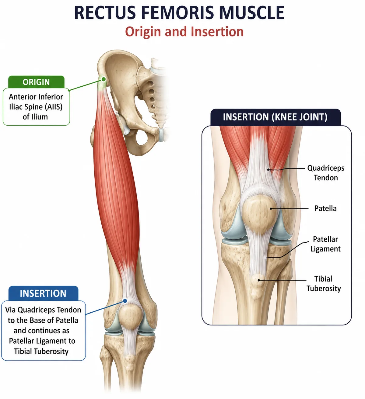
Origin
- Anterior inferior iliac spine (AIIS)
- Area above the acetabulum
Insertion
- Patella through quadriceps tendon
- Tibial tuberosity through patellar ligament
Function
- Extends the knee.
- Assists hip flexion.
Clinical Importance
Because it crosses both the hip and knee, rectus femoris is a biarticular muscle.
B. Vastus Lateralis
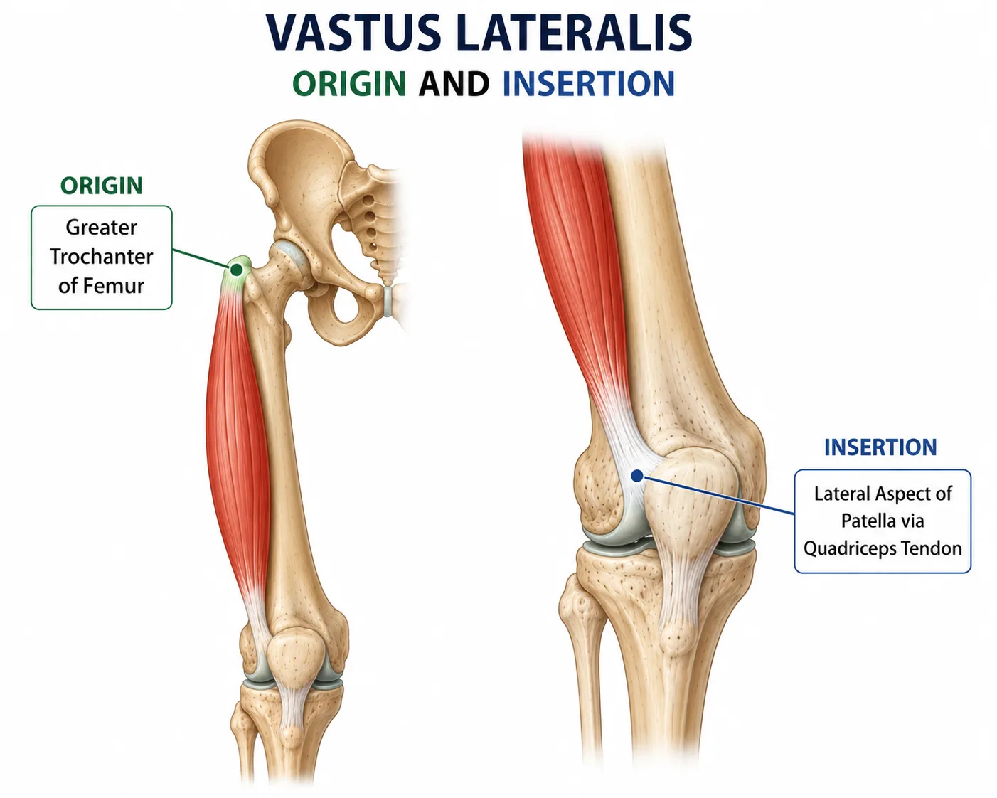
Origin
- Greater trochanter
- Lateral lip of linea aspera
- Lateral femur
Insertion
- Patella through quadriceps tendon
- Tibial tuberosity through patellar ligament
Function
- Extends the knee.
- Helps stabilize the patella.
C. Vastus Medialis
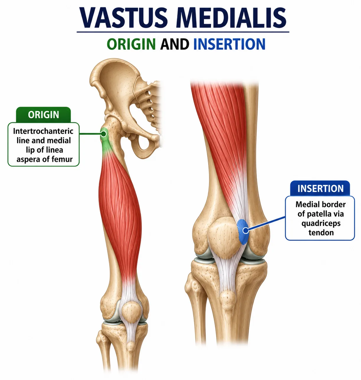
Origin
- Medial femur
- Medial lip of linea aspera
Insertion
- Patella through quadriceps tendon
- Tibial tuberosity through patellar ligament
Function
- Extends the knee.
- Helps maintain medial patellar stability.
Important Part: VMO
The vastus medialis obliquus (VMO) is an important portion of the vastus medialis that contributes to medial control of the patella.
D. Vastus Intermedius
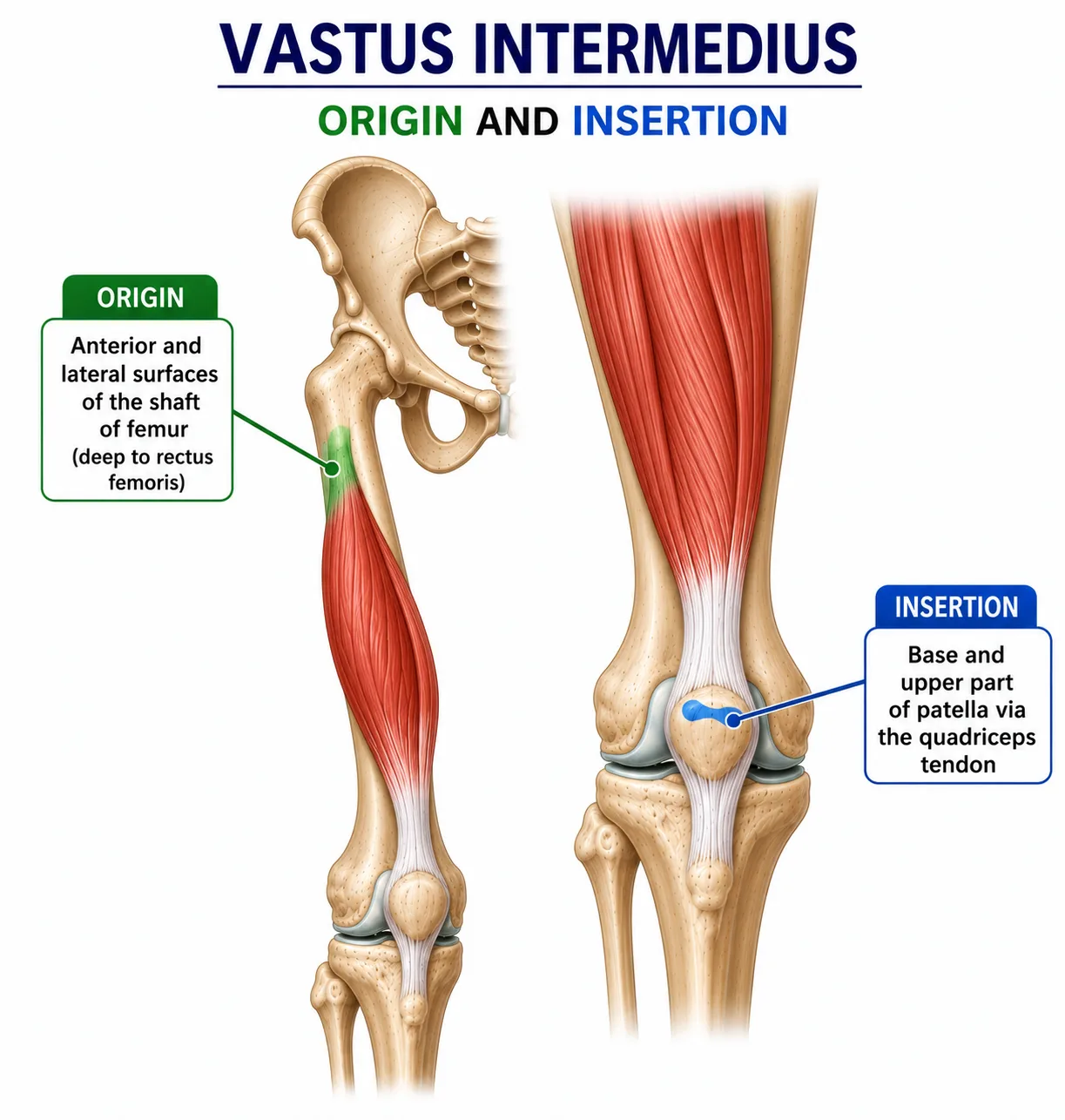
Origin
- Anterior and lateral surfaces of the shaft of the femur
Insertion
- Patella through quadriceps tendon
- Tibial tuberosity through patellar ligament
Function
- Extends the knee.
6. Hamstring Muscles
The hamstrings are located on the posterior aspect of the thigh.
The three main hamstrings are:
- Biceps femoris
- Semitendinosus
- Semimembranosus
Their major action at the knee is flexion.
A. Biceps Femoris
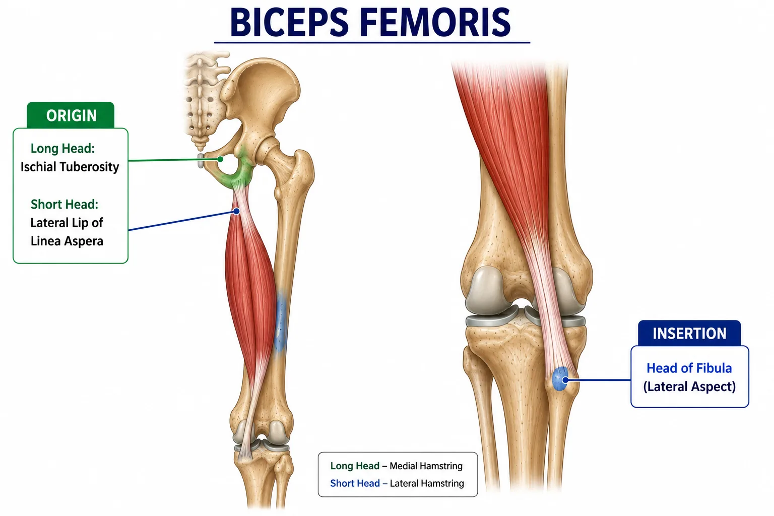
It has two heads:
Long Head Origin
- Ischial tuberosity
Short Head Origin
- Linea aspera
- Lateral supracondylar line of femur
Insertion
- Head of fibula
Function
- Flexes the knee.
- Laterally rotates the leg when the knee is flexed.
- Long head also assists hip extension.
B. Semitendinosus
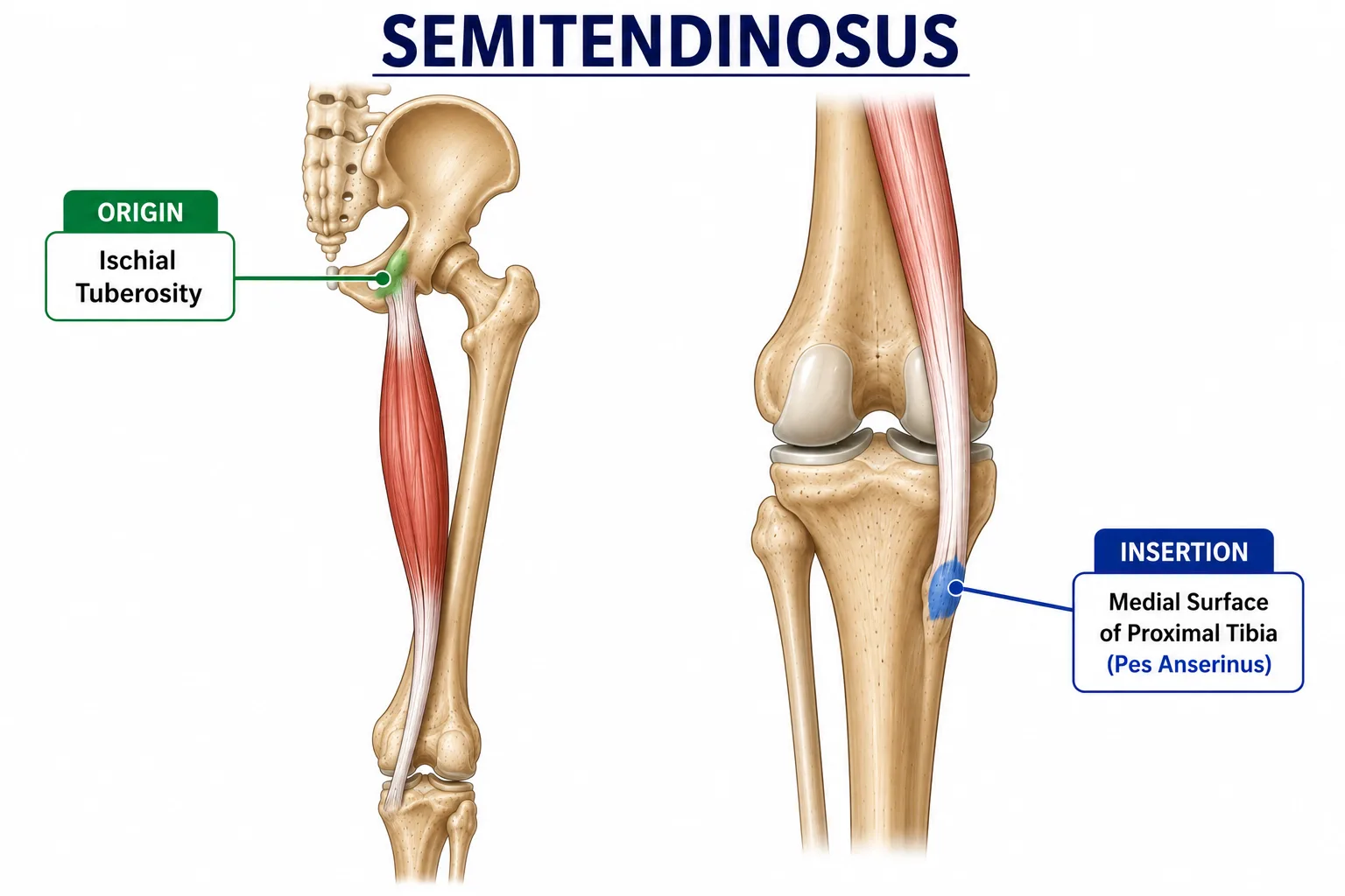
Origin
- Ischial tuberosity
Insertion
- Medial surface of proximal tibia
- Part of the pes anserinus
Function
- Flexes the knee.
- Medially rotates the leg when the knee is flexed.
- Assists hip extension.
C. Semimembranosus
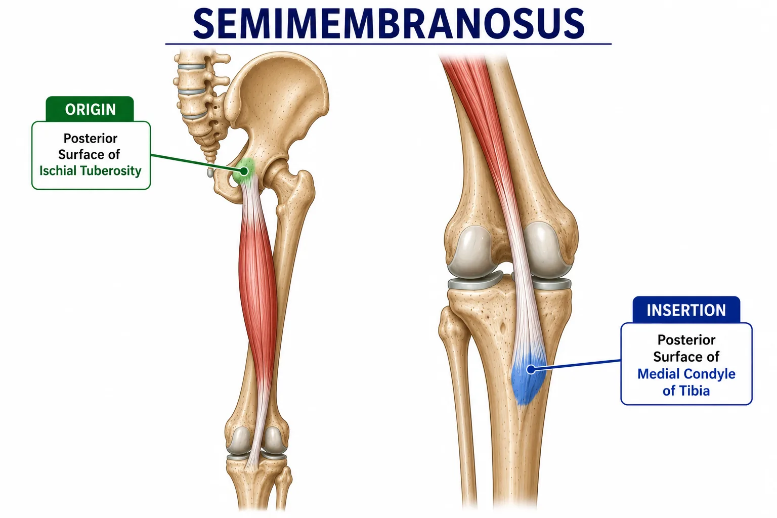
Origin
- Ischial tuberosity
Insertion
- Posteromedial surface of proximal tibia
Function
- Flexes the knee.
- Medially rotates the leg when the knee is flexed.
- Assists hip extension.
7. Sartorius
The sartorius is the longest muscle in the human body.
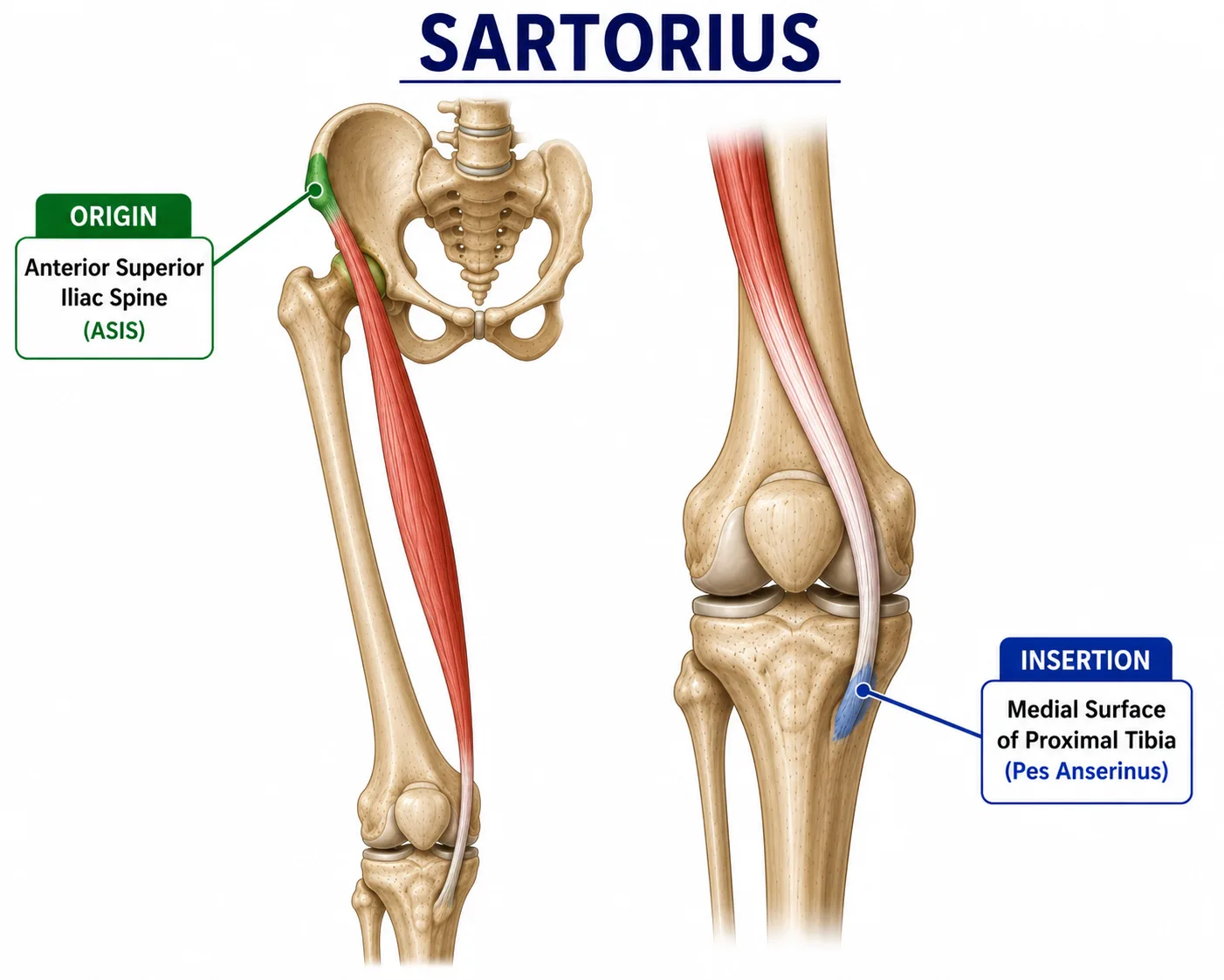
Origin
- Anterior superior iliac spine (ASIS)
Insertion
- Medial surface of proximal tibia through the pes anserinus
Function
- Flexes the knee.
- Medially rotates the leg when the knee is flexed.
- Also assists hip flexion and lateral rotation.
Simple Explanation
Sartorius helps produce the movement used when sitting cross-legged.
8. Gracilis
The gracilis is a long, thin muscle on the medial side of the thigh.
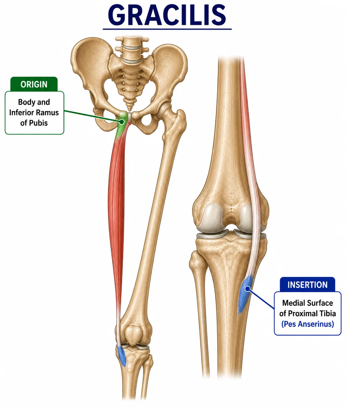
Origin
- Inferior pubic ramus
- Body of pubis
Insertion
- Medial proximal tibia through the pes anserinus
Function
- Adducts the hip.
- Flexes the knee.
- Medially rotates the leg when the knee is flexed.
9. Gastrocnemius
The gastrocnemius is the large calf muscle that crosses the knee joint.
It has two heads.
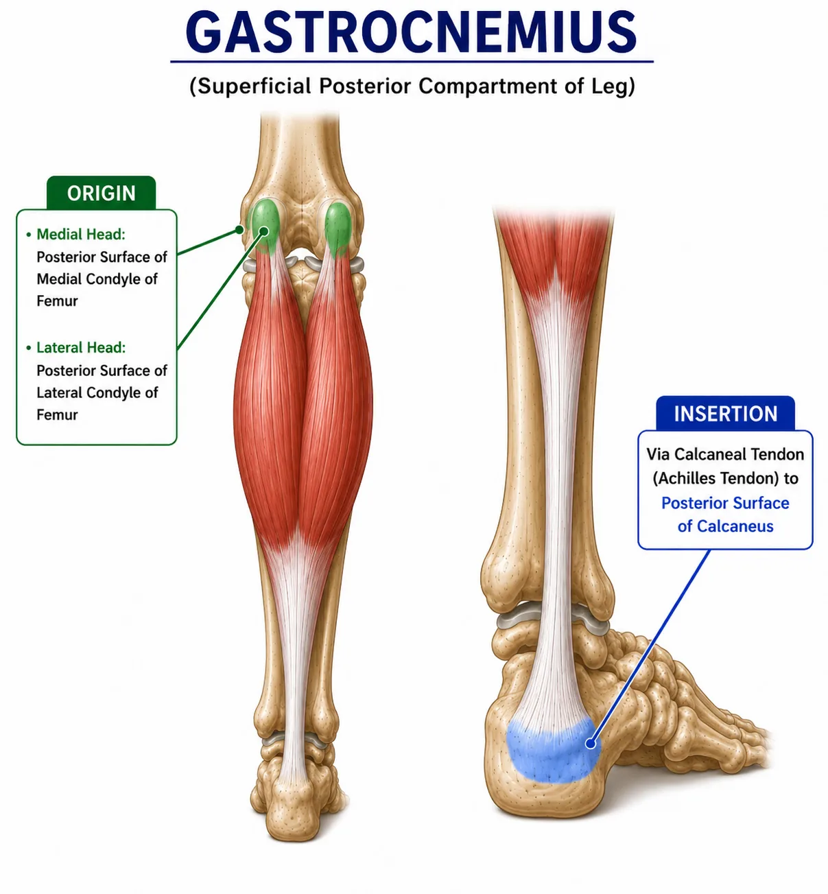
Origin
- Medial femoral condyle
- Lateral femoral condyle
Insertion
- Calcaneus through the Achilles tendon
Function
- Assists knee flexion.
- Produces plantarflexion of the ankle.
Clinical Importance
Because gastrocnemius crosses both the knee and ankle, its length and flexibility can influence movement at both joints.
10. Popliteus
The popliteus is a small but important muscle located at the back of the knee.
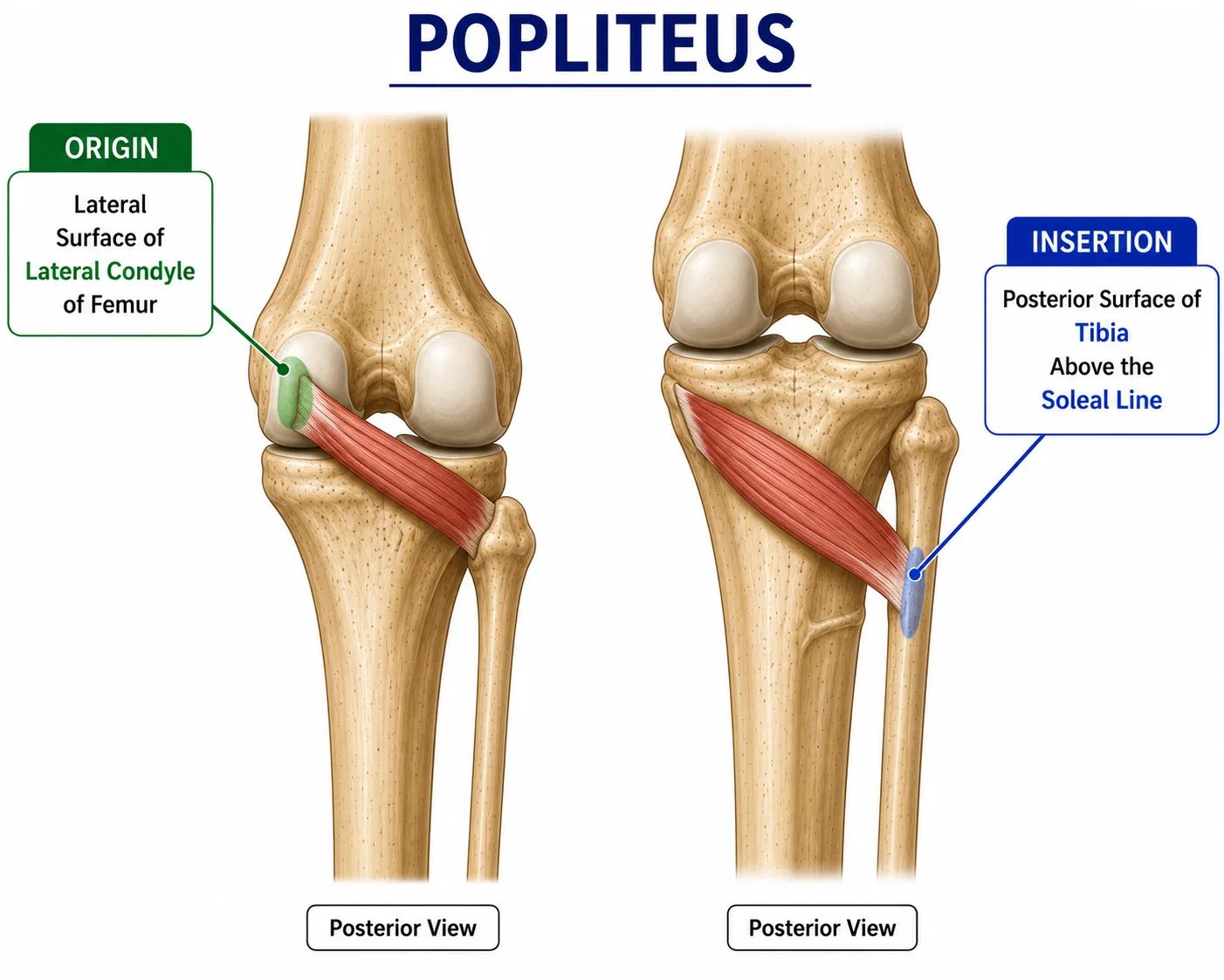
Origin
- Lateral femoral condyle
Insertion
- Posterior surface of proximal tibia
Function
- Helps initiate knee flexion.
- Medially rotates the tibia when the foot is free.
- Helps laterally rotate the femur when the foot is fixed.
- Helps unlock the knee from full extension.
Simple Explanation
The popliteus helps “unlock” the knee when beginning to bend it from a fully straight position.
11. Tensor Fasciae Latae and Iliotibial Tract
The tensor fasciae latae does not directly insert on the tibia as a muscle, but its force is transmitted through the iliotibial tract (IT band).
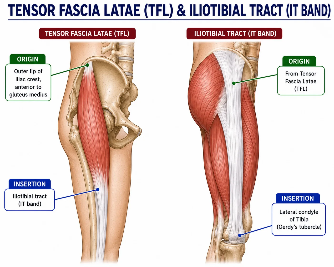
Origin
- Anterior part of iliac crest
- ASIS region
Insertion
- Lateral condyle of tibia through the iliotibial tract
Function
- Helps stabilize the knee during standing and movement.
- Assists knee extension when the knee is near full extension.
- Helps maintain lateral stability of the knee.
12. Quick Revision Table
| Structure | Origin | Insertion | Main Function |
| ACL | Tibia | Femur | Prevents excessive anterior tibial translation |
| PCL | Tibia | Femur | Prevents excessive posterior tibial translation |
| MCL | Medial femur | Medial tibia | Resists valgus stress |
| LCL | Lateral femur | Fibula | Resists varus stress |
| Rectus femoris | AIIS | Patella → tibial tuberosity | Knee extension + hip flexion |
| Vastus lateralis | Lateral femur | Patella → tibial tuberosity | Knee extension |
| Vastus medialis | Medial femur | Patella → tibial tuberosity | Knee extension + patellar stability |
| Vastus intermedius | Femur | Patella → tibial tuberosity | Knee extension |
| Biceps femoris | Ischial tuberosity/femur | Fibular head | Knee flexion + lateral rotation |
| Semitendinosus | Ischial tuberosity | Medial tibia | Knee flexion + medial rotation |
| Semimembranosus | Ischial tuberosity | Posteromedial tibia | Knee flexion + medial rotation |
| Sartorius | ASIS | Medial tibia | Knee flexion + medial rotation |
| Gracilis | Pubis | Medial tibia | Knee flexion + medial rotation |
| Gastrocnemius | Femoral condyles | Calcaneus | Knee flexion + plantarflexion |
| Popliteus | Lateral femoral condyle | Posterior tibia | Unlocks knee |
| TFL/IT tract | Iliac region | Lateral tibia | Lateral knee stability |
13. Knee Movements and Main Muscles
Knee Extension
Main muscle:
Quadriceps femoris
Knee Flexion
Main muscles:
Hamstrings
Assist:
- Gastrocnemius
- Sartorius
- Gracilis
- Popliteus
Medial Rotation of Tibia
Main contributors:
- Semitendinosus
- Semimembranosus
- Sartorius
- Gracilis
- Popliteus
Lateral Rotation of Tibia
Main contributor:
- Biceps femoris
14. Why Knee Stability Is Important
Knee stability comes from a combination of:
Bones + Ligaments + Menisci + Muscles + Joint Capsule
Ligaments provide passive stability, while muscles provide dynamic stability.
For example:
- The ACL controls excessive anterior tibial movement.
- The PCL controls excessive posterior tibial movement.
- The MCL protects against valgus stress.
- The LCL protects against varus stress.
- The quadriceps and hamstrings provide dynamic control during walking, running and sports.
Good coordination between these structures is essential for normal knee function.
Conclusion
The knee is a complex joint that depends on several structures working together. The ACL, PCL, MCL and LCL provide important ligamentous stability, while the quadriceps, hamstrings, gastrocnemius, sartorius, gracilis and popliteus control movement and provide dynamic support.
Understanding the origin, insertion and function of these muscles and the role of each ligament is essential for physiotherapy students, clinicians, athletes and anyone interested in knee health.
This anatomical knowledge also helps in understanding common knee conditions such as ACL injury, meniscus injury, MCL/LCL injury, patellofemoral pain, osteoarthritis and sports-related knee injuries.
Educational Note: This information is intended for anatomy and physiotherapy education. Individual knee injuries and rehabilitation should be assessed by an appropriately qualified healthcare professional.